Chronic lower limb ulcers are a growing concern in Kenya, especially among adults with underlying vascular conditions. Among the most prevalent types are arterial ulcers and venous ulcers, each with distinct causes, clinical features, and management protocols. Early diagnosis and specialized wound care significantly improve outcomes and reduce the risk of amputation.

This article offers an in-depth comparison of arterial and venous ulcers and provides a clinical guide for patients, caregivers, and healthcare professionals seeking effective vascular wound care in Nairobi and beyond.
What Are Arterial Ulcers?
Arterial ulcers, also known as ischemic ulcers, result from insufficient blood flow to the lower extremities. They often affect patients with peripheral arterial disease (PAD), diabetes mellitus, or advanced atherosclerosis.

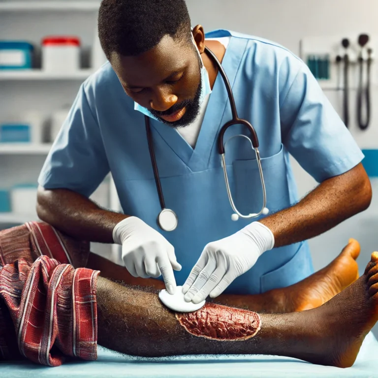
a mixture of arterial ulcer and a venous ulcer
Clinical Features:
- Located on the toes, heels, or pressure points of the foot
- Ulcers appear punched out, with well-defined edges
- Dry, necrotic base, often covered with black eschar
- Surrounding skin is cool, pale, or cyanotic
- Severe pain, especially at night or when the legs are elevated
- Weak or absent peripheral pulses
Causes and Risk Factors:
- Peripheral arterial disease (PAD)
- Diabetes mellitus
- Smoking
- Hypertension
- Hyperlipidemia
- Advanced age
Diagnosis:
A comprehensive vascular assessment includes:
- Ankle-brachial index (ABI)
- Doppler ultrasound
- Toe pressures and transcutaneous oxygen measurements (TcPO2)
What Are Venous Ulcers?
Venous ulcers, also known as stasis ulcers, occur due to chronic venous insufficiency (CVI) and venous hypertension. These are the most common type of chronic leg ulcers and typically affect the medial gaiter area of the lower leg.

Clinical Features:
- Shallow, irregular-shaped ulcers with granulating base
- Surrounding skin shows hyperpigmentation, lipodermatosclerosis, and varicosities
- Minimal pain, often relieved by leg elevation
- Edema of the lower limbs
- Warm skin with normal peripheral pulses
Causes and Risk Factors:
- Deep vein thrombosis (DVT)
- Varicose veins
- Obesity
- Pregnancy
- Sedentary lifestyle
- Long-standing CVI
Diagnosis:
- Venous Doppler ultrasound to assess reflux and obstruction
- Clinical history and physical exam
- Duplex imaging
Key Differences Between Arterial and Venous Ulcers
| Feature | Arterial Ulcers | Venous Ulcers |
| Location | Toes, heels, pressure points | Medial malleolus (gaiter area) |
| Pain | Severe, nocturna | Mild to moderate |
| Appearance | Punched-out, necrotic | Irregular, shallow, granulating |
| Skin Condition | Cold, hairless, shiny | Warm, edematous, discolored |
| Pulses | Weak or absent |
Present |
| Response to Elevation | Worsens pain | Relieves symptoms |
Wound Management Strategies
Arterial Ulcer Management:
- Revascularization is key: angioplasty or bypass surgery
- Avoid compression therapy
- Maintain dry, sterile dressings
- Optimize underlying conditions: control blood glucose, hypertension, and cholesterol
- Smoking cessation and exercise therapy when appropriate
- Pain control and infection surveillance
Venous Ulcer Management:
- Graduated compression therapy (with ABPI confirmation >0.8)
- Elevation of legs
- Regular wound cleansing and moist wound healing techniques
- Topical dressings (e.g., foam, hydrocolloids)
- Long-term venous reflux management, including ablation or surgery if needed
Advanced Wound Care Options in Nairobi
At VitalCare Wound & Ostomy Care, we specialize in the comprehensive evaluation and treatment of complex wounds, including arterial and venous ulcers. Our services include:
- Vascular assessment tools: ABI, Doppler, and capillary refill analysis
- Advanced wound therapies: negative pressure wound therapy (VAC), debridement, and biologic dressings
- Customized compression therapy
- Patient education on lifestyle changes and ulcer prevention
- Multidisciplinary care including collaboration with vascular surgeons, endocrinologists, and physiotherapists
Prevention and Patient Education
Preventive strategies significantly reduce the recurrence and progression of chronic vascular ulcers:
- Monitor and manage chronic illnesses like diabetes and hypertension
- Promote daily skin inspection and foot care
- Encourage mobility and leg elevation
- Use proper footwear to reduce trauma
- Ensure adherence to compression garment protocols for venous disease
When to Seek Specialized Wound Care
Early intervention saves limbs. Seek expert wound care if:
- The ulcer has been present for more than 2–3 weeks without healing
- You observe increasing pain, swelling, or discharge
- There are signs of infection (redness, warmth, pus)
- There is skin discolouration, gangrene, or non-palpable pulses
Conclusion
Arterial and venous ulcers require distinct diagnostic and treatment approaches, yet both benefit from early detection, multidisciplinary care, and individualized wound management plans. At Stanley Wound & Ostomy Care in Nairobi, we are committed to offering evidence-based solutions for complex wound conditions across Kenya.
If you or a loved one is living with a chronic leg ulcer, don’t wait. Contact us today for a consultation and access to expert wound care.
Book an Appointment Today
📍 Nairobi, Kenya
📞 +254 701287584
🌐 VitalCare – VitalCare Wound and Ostomy clinic
📧 admin@woundandostomycare.co.ke
VitalCare Wound and Ostomy clinic – Restoring Health, One Wound at a Time.